We Finally Know Why There's a Bizarre Structure in Our Inner Ears
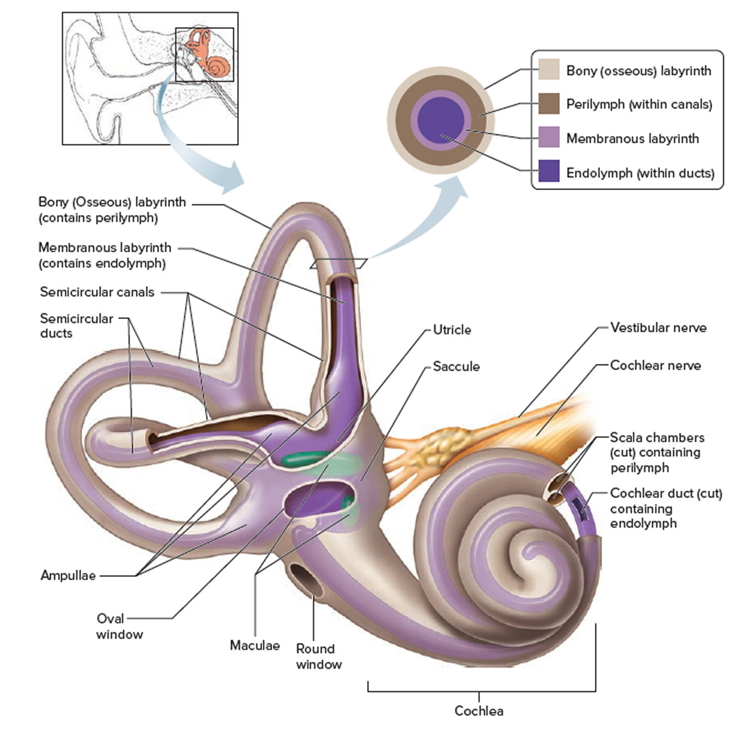
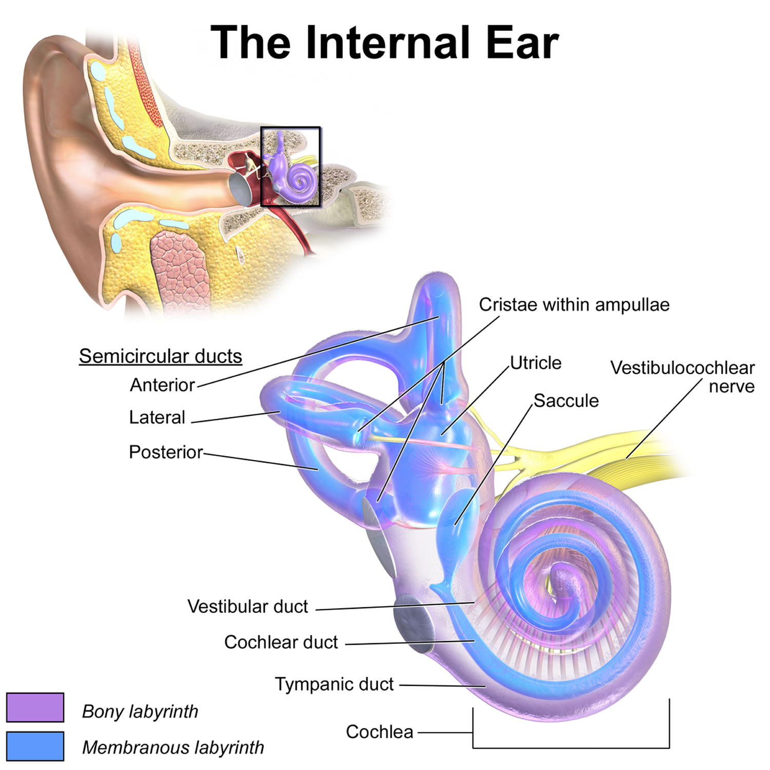
inner ear, part of the ear that contains organs of the senses of hearing and equilibrium. The bony labyrinth, a cavity in the temporal bone, is divided into three sections: the vestibule, the semicircular canals, and the cochlea. Within the bony labyrinth is a membranous labyrinth, which is also divided into three parts: the semicircular ducts.
SPEECH LANGUAGE PATHOLOGY & AUDIOLOGY HEARING DISORDERS OF THE OUTER EAR
Anatomy Function Associated Conditions Tests Essential for hearing and balance, each ear has an intricate structure of bones, nerves, and muscles. The ears can be affected by bacterial infections, viral infections, hearing loss, tinnitus (ringing in the ears), Meniere's disease, and more. Anatomy

EarQ Anatomy of the Ear Chart Human ear, Inner ear diagram, Ear anatomy
Anatomy What are the parts of the inner ear? Your inner ear has three main parts: your cochlea, semi-circular canals (labyrinth) and your vestibule. Your cochlea supports your hearing and your vestibule and semi-circular canals support your balance. What is the cochlea?
Ear infections explained Dr Mark McGrath
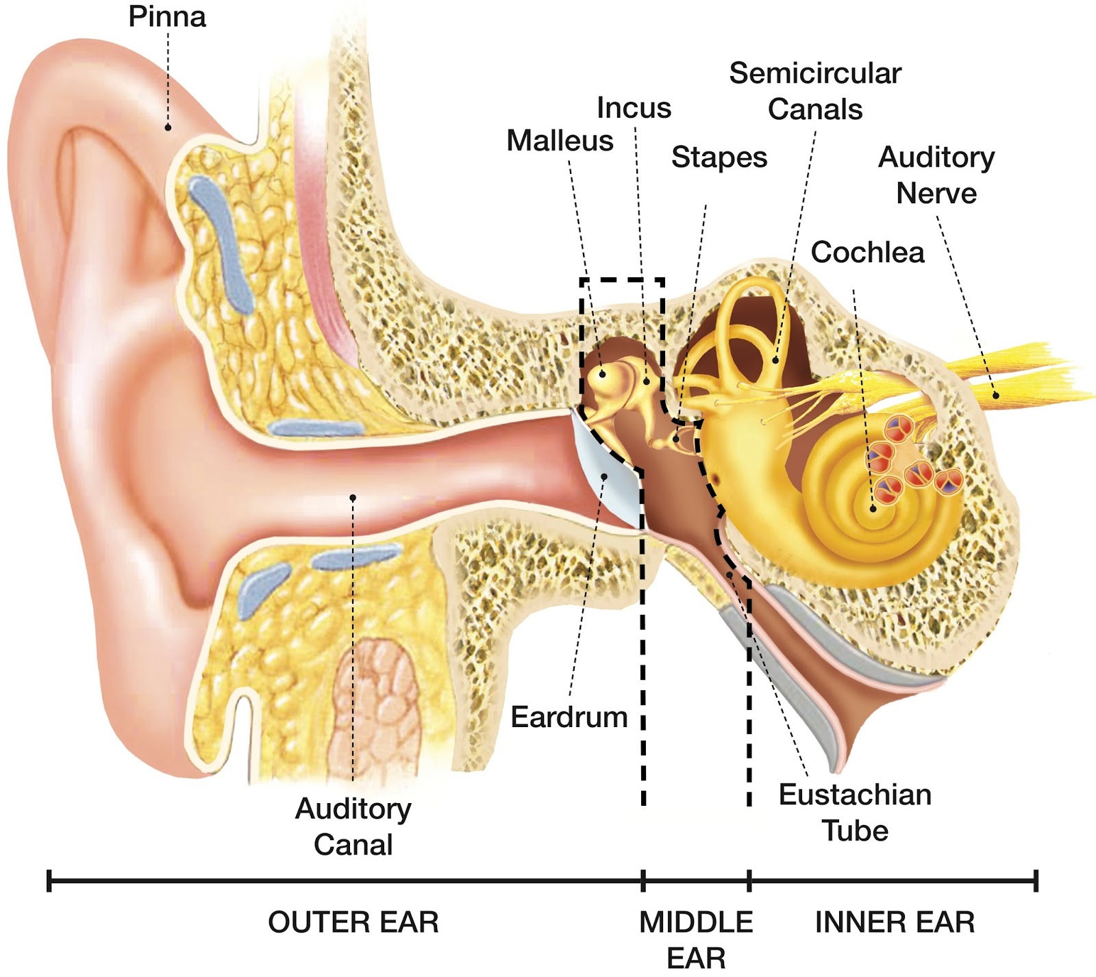
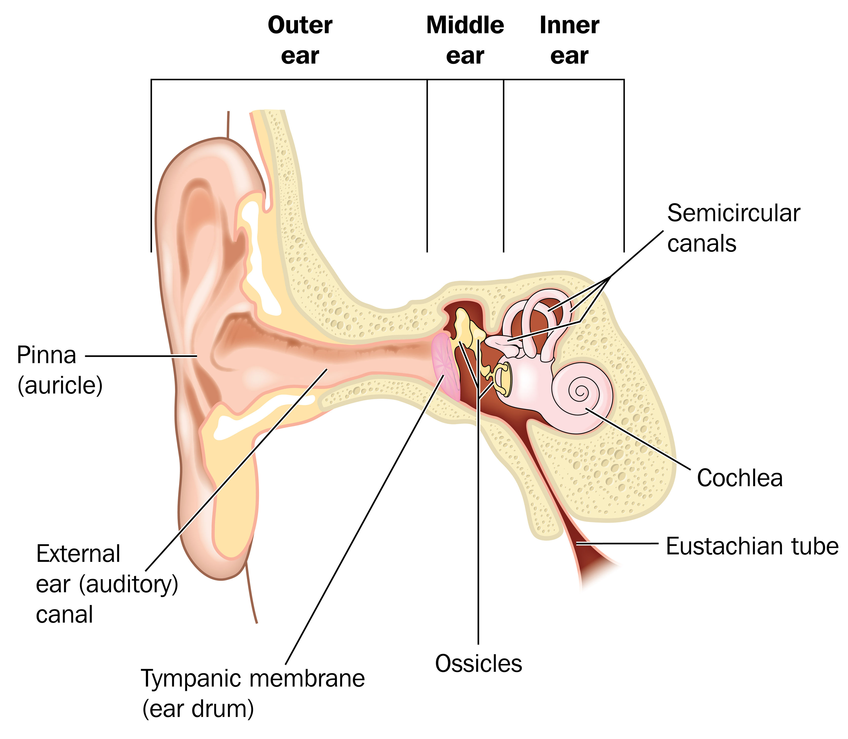
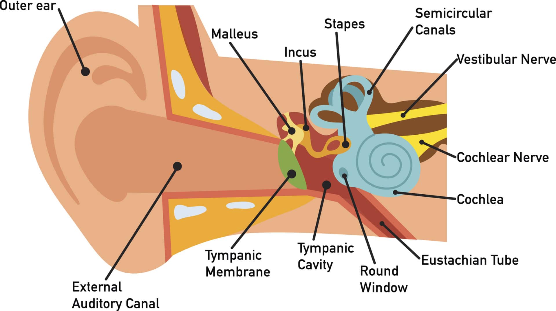
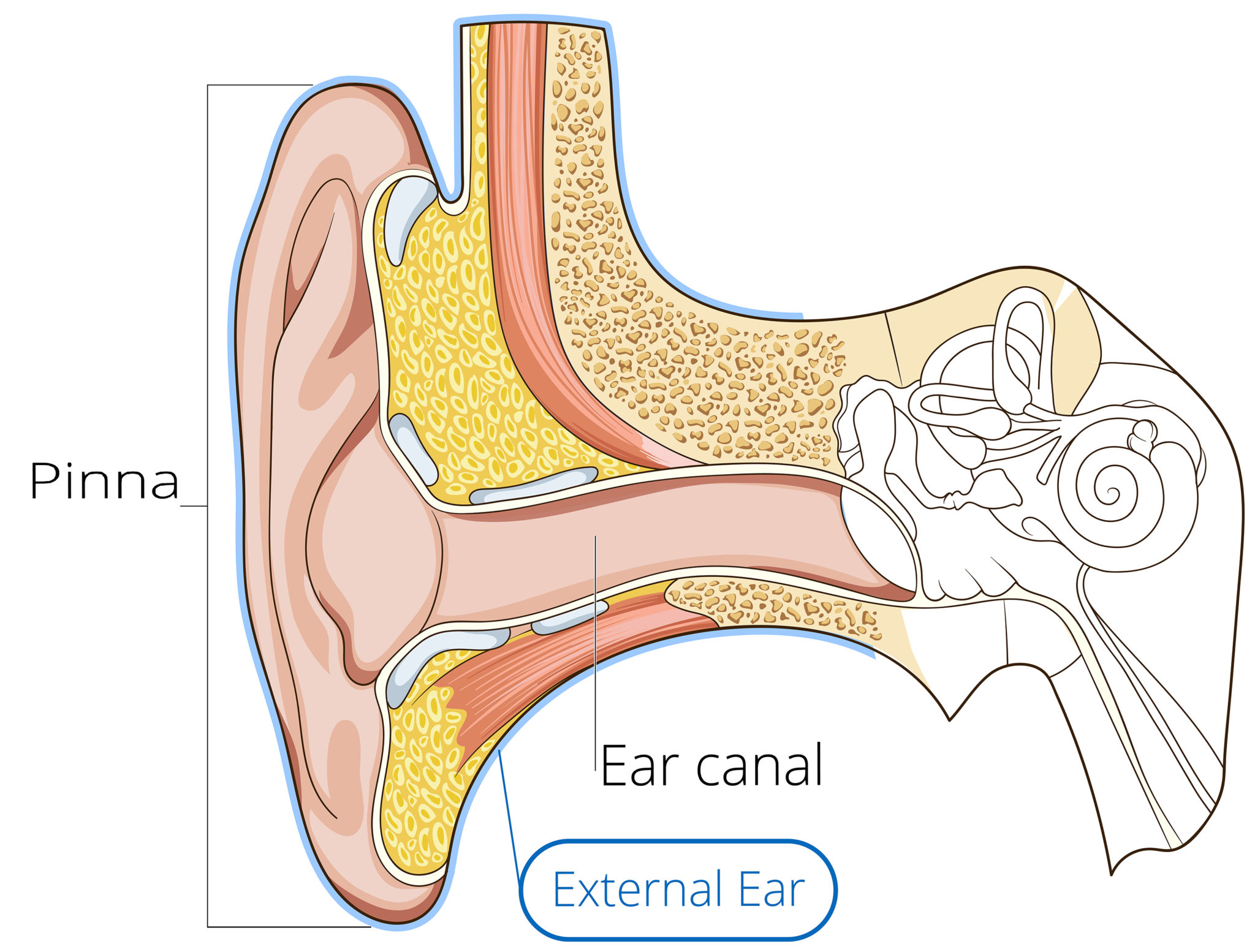
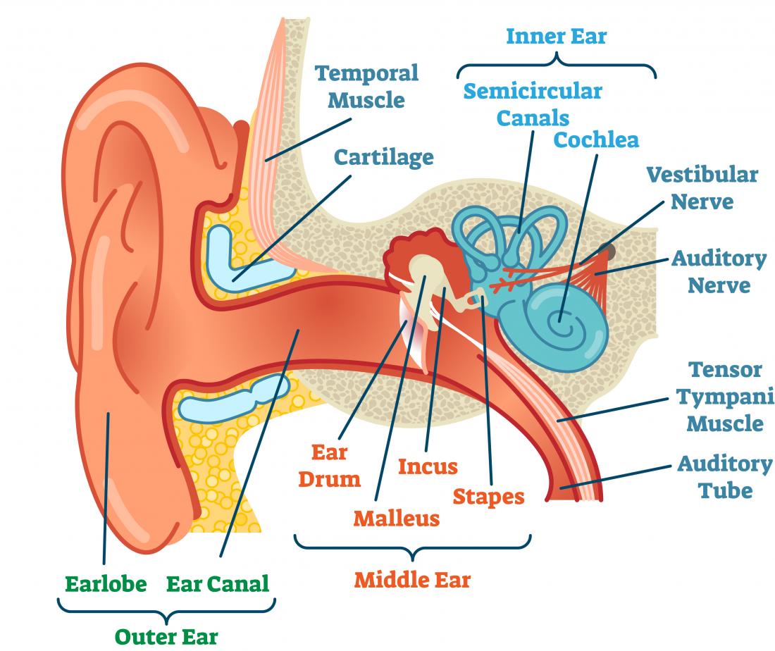
Click Image to Enlarge. The ear is the organ of hearing and balance. The parts of the ear include: External or outer ear, consisting of: Pinna or auricle. This is the outside part of the ear. External auditory canal or tube. This is the tube that connects the outer ear to the inside or middle ear. Tympanic membrane (eardrum).

Hearing Physics
1/4 Synonyms: External auditory meatus, External acoustic pore , show more. The ear is a complex part of an even more complex sensory system. It is situated bilaterally on the human skull, at the same level as the nose. The main functions of the ear are, of course, hearing, as well as constantly maintaining balance.
How does your ear work?
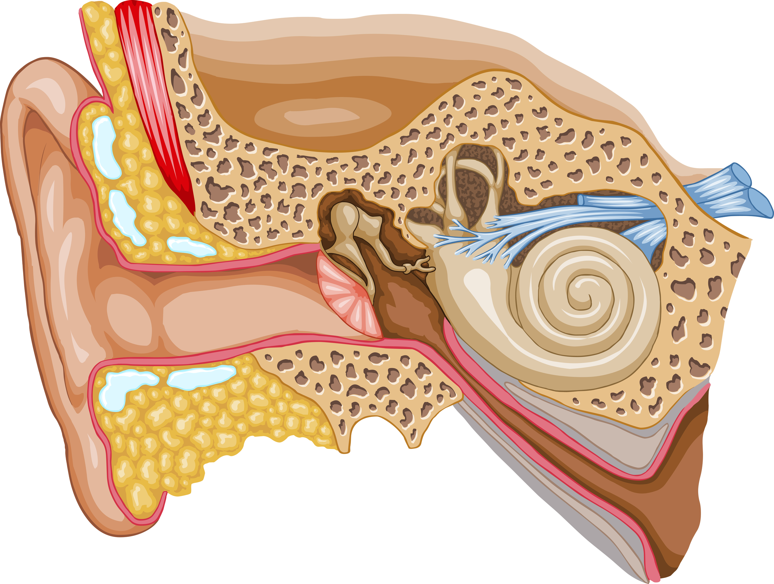
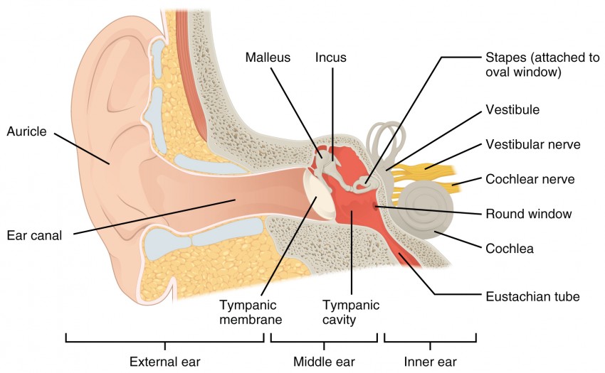
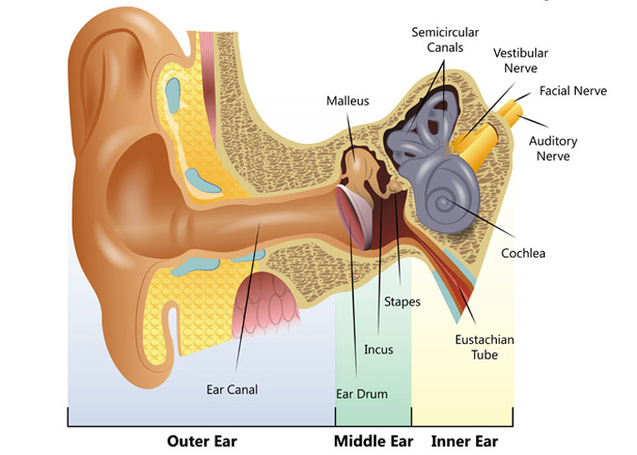
Structure and Function. The ear is organized into three different anatomical structures: the outer, middle, and inner ear. The outer ear consists of the pinna, external auditory canal, and tympanic membrane and is responsible for the transmission of sound waves from the external environment. The middle ear is an air-filled space that contains the three ossicles (malleus, incus, and stapes.
How You Hear Northland Audiology
So as the air vibrates even the ear drum starts vibrating. Just like the skin of a drum. And as you can, the ear drum also separates the outer ear from the middle ear. This brings us to the middle ear. The middle ear consists of the three tiniest bones of the human body. And they're together the are called the ossicles. And they have pretty.
Medical problems of the eyes, ears, nose, and throat symptoms
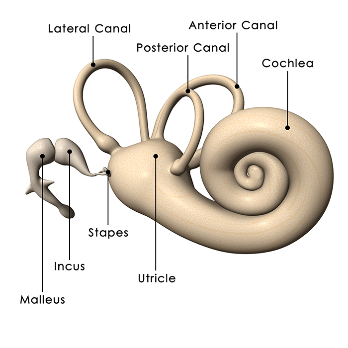
Structure The ear is made up of the outer ear, middle ear, and inner ear. The inner ear consists of the bony labyrinth and membranous labyrinth. The bony labyrinth comprises three components: Cochlea: The cochlea is made of a hollow bone shaped like a snail and divided into two chambers by a membrane.
Ear Anatomy Causes of Hearing Loss Hearing Aids Audiology
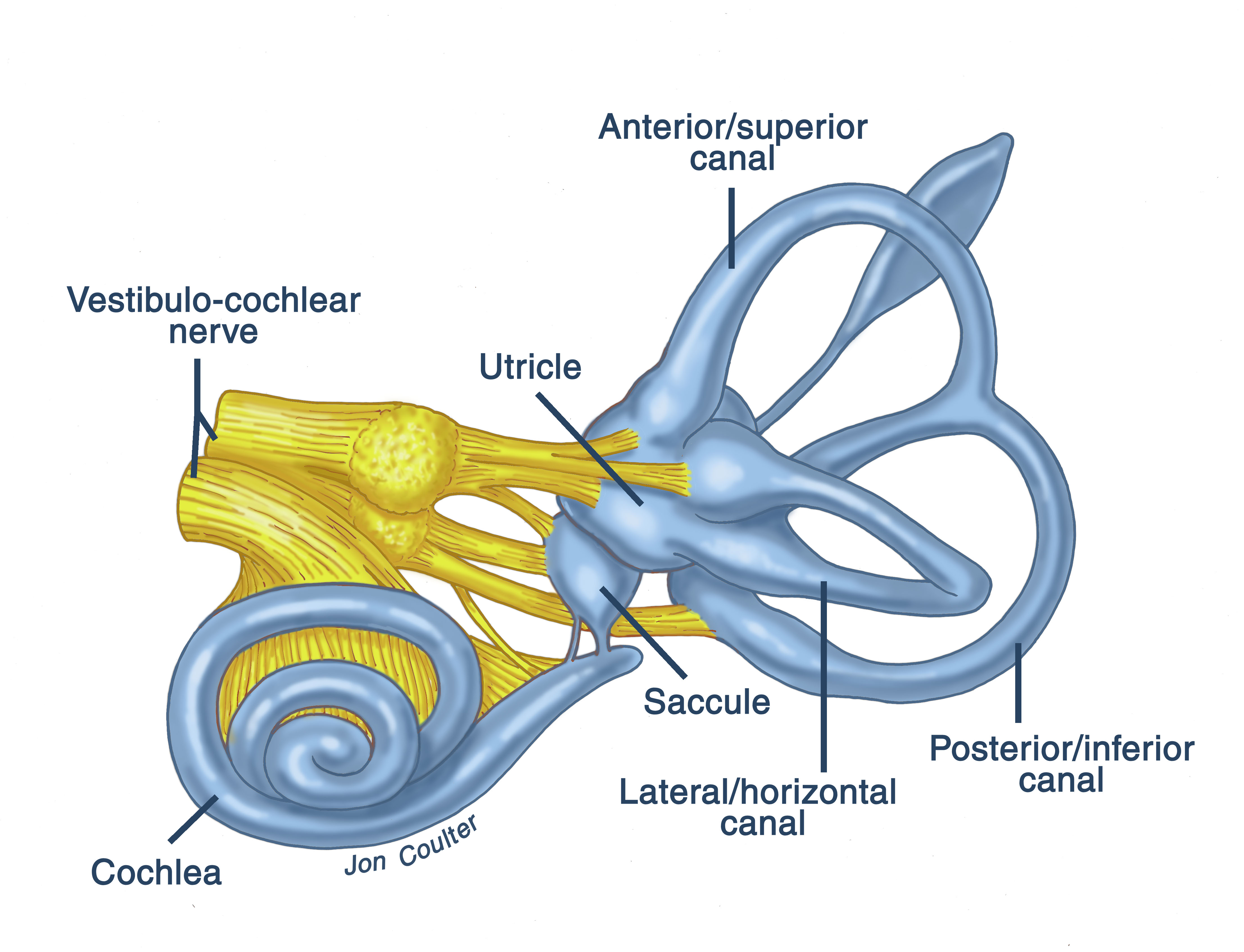
Balance: Your inner ear contains semicircular canals filled with fluid and hair-like sensors. When you move your head, the fluid inside these loop-shaped canals sloshes around and moves the hairs. The hairs transmit this information along the vestibular nerve to your brain.
Inner Ear Problems Causes & Treatment of inner ear Dizziness & Vertigo
The outer ear consists of the visible portion called the auricle, or pinna, which projects from the side of the head, and the short external auditory canal, the inner end of which is closed by the tympanic membrane, commonly called the eardrum. The function of the outer ear is to collect sound waves and guide them to the tympanic membrane.
How The Ear Works Step by Step Brief Explanation
The inner ear is embedded within the petrous part of the temporal bone, anterolateral to the posterior cranial fossa, with the medial wall of the middle ear, the promontory, serving as its lateral wall.

Disorders of the Ear Part Two a PA Review and Podcast
The hearing part of the inner ear is called the cochlea. This comes from the Greek word for 'snail' because of its distinctive coiled shape. The cochlea, which contains many thousands of sensory cells (called 'hair cells'), is connected to the central hearing system by the hearing or auditory nerve.

Human ear anatomy. Ears inner structure, organ of hearing ve (1000410
Inner ear: The inner ear, also called the labyrinth, operates the body's sense of balance and contains the hearing organ. A bony casing houses a complex system of membranous cells. The.
Audition and Somatosensation Anatomy and Physiology I
Sound waves are created when air vibrates. To hear, the ear needs to change sound into electrical signals which the brain can interpret. The outer part of the ear (the pinna) funnels sound waves into the ear canal. When sound waves reach the eardrum they cause it to vibrate. Vibrations of the eardrum cause the tiny bones in the middle ear to.
Common balance disorders Hearing Link
Anatomy. The inner ear is a complex three dimensional shape with semicircular canals, dilations called the utricle and saccule and a spiral portion known as the cochlea. All of these organs are housed inside a bony shell known as the bony labyrinth and this is within the temporal bone. The cochlea is the site where sound is transformed into.
Inner Ear Problems Causes & Treatment of inner ear Dizziness & Vertigo
The ear can be divided into three parts: the external, middle and inner ear. The ears are an organ of hearing and balance, converting information from our external environment into electrical signals that can be processed by the brain. This article will explore the three anatomical sections of the ear, highlighting their individual anatomy and.