Panuveitis
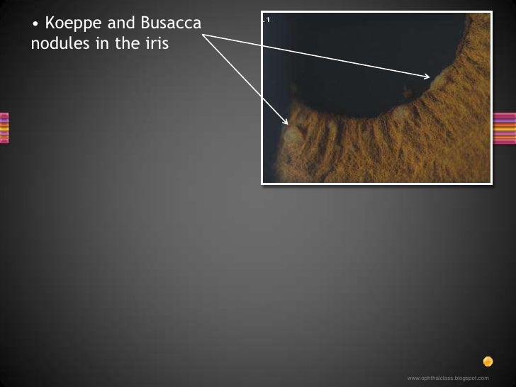
Other features of granulomatous anterior uveitis include presence of Koeppe's nodules at the pupillary border and/or Busacca's nodules in the iris stroma.

Iris nodules ️Koeppe... OphthalmologyNotes And Synopses Facebook
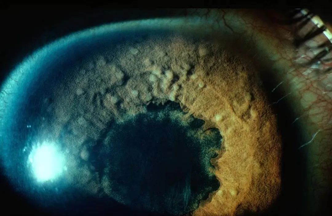
Koeppe nodules Condition/keywords acute anterior uveitis Photographer Divya Jain Imaging device 35mm external camera Description Slit Lamp photograph of a 33 year old woman with first episode of acute granulomatous anterior uveitis showing mutton fat KP'S and Koeppe's nodules. Related files

Multiple Koeppe nodules (white arrows) at iris pupillary border noted
Koeppe's Nodules May be found as: Koeppe nodules (inflammatory cell precipitates which lie at the pupillary margin and could be found in non-granulomaous as well as granulomatous uveitis) Bussaca nodules (lie on the iris surface) which are pathognomonic for granulomatous uveitis. When the inflammation is treated, the nodules will resolve.

lisch nodules Google Search NF 1 Анатомия человека, Искусство глаза
The Standardization of Uveitis Nomenclature (SUN) Working Group classifies onset as either "sudden," which is characterized by pain, redness and photophobia, or "insidious," where the eye is painless and white.2 • Duration.
肉芽肿性炎和非肉芽肿性炎有何区别?4个结节要分清_医学界助力医生临床决策和职业成长
In comparison, smaller koeppe nodules located on the pupillary border may be seen in both granulomatous and nongranulomatous irides. 7,8 Posterior segment involvement occurs in approximately one third of patients with ocular sarcoidosis, and typically presents as an intermediate uveitis. 3,7,8 Peripheral neovascularization, with or without.
Koeppe nodules and muttonfat keratic precipitates American Academy
Objectives: Describe the pathogenesis of ocular sarcoidosis. Summarize the relevant history of patients with ocular sarcoidosis. Review the evaluation process of patients with ocular sarcoidosis. Outline the management of ocular sarcoidosis, including the role of interprofessional care of these patients.
Atlas Entry Sarcoidosis
Viral anterior uveitis (VAU) is characterized by anterior uveitis (AU) with elevated intraocular pressure (IOP) diffuse stellate keratic precipitates (KPs), presence of pigmentation in active KPs and iris atrophic changes. [ 1 2 3 4] The most commonly implicated viruses include herpes simplex (HSV), varicella-zoster (VZV), cytomegalovirus (CMV),.

Iris nodules in Fuchs heterochromic iridocyclitis. Abstract Europe PMC
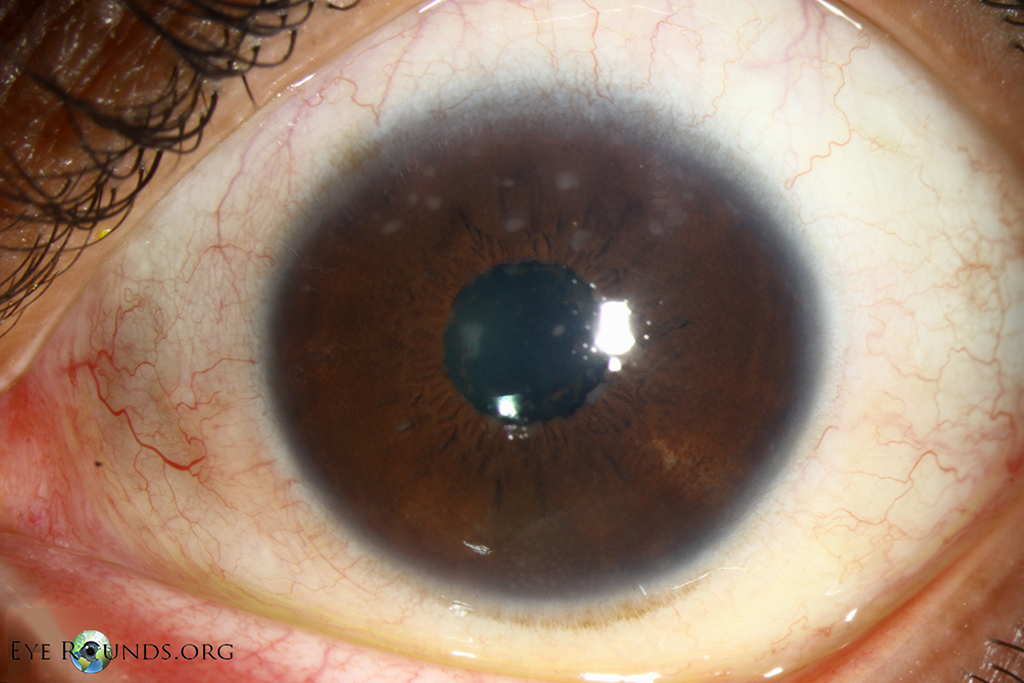
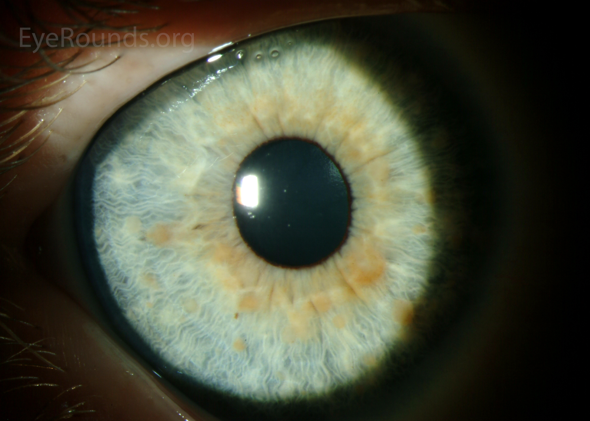
iris nodules: Koeppe (small, near pupil), Bussaca (large, far from pupil) Anterior vitreous cells may be seen in iridocyclitis but often will indicate intermediate ± posterior uveitis Other signs include constricted or non-reactive pupil, iris atrophy, heterochromia, cataract, chronic corneal oedema including bullous keratopathy
Atlas Entry Lisch nodules
Sarcoid Uveitis. by Edmund Tsui, MD on September 19, 2023. Sarcoidosis is a systemic inflammatory disease characterized by the formation of noncaseating granulomas in affected organs, most commonly the lungs, lymph nodes, skin, and eyes. The disease was first described in 1878 by noted surgeon Sir Jonathan Hutchinson as a dermatologic disorder [1].

NODULES in Ophthalmology
Sarcoidosis is a systemic inflammatory disease of unknown etiology characterized by the formation of noncaseating granulomas. The disease most commonly affects the skin, lungs, lymph nodes, and eyes but can affect virtually any organ.
Koeppe nodules American Academy of Ophthalmology
The diseases most commonly associated with iris nodules and uveitis include sarcoidosis, Vogt-Koyanagi-Harada syndrome, multiple sclerosis, Fuchs' heterochromic iridocyclitis, and metastatic infection.

Iris nodules ️Koeppe... OphthalmologyNotes And Synopses Facebook
Nodules found at the pupillary border are known as Koeppe nodules and those found on the surface of the iris are referred to as Busaca nodules. 7. FUS is a clinical diagnosis, dependent on the eye examination. To detect the presence of rubella-specific antibodies, an anterior chamber paracentesis may be completed, however, this is not a.

Pupil margin (Koeppe) and iris surface (Busacca) nodules in a patient
Koeppe's nodules are small nodules seen at the inner margin of the iris in patients with granulomatous anterior uveitis, which occurs in conditions such as sarcoidosis and tuberculosis. The nodules are composed of epithelioid cells and giant cells surrounded by lymphocytes.
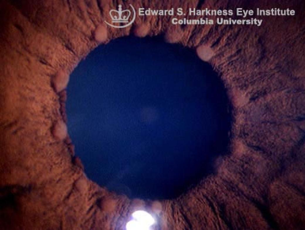
Koeppe's Nodules Vagelos College of Physicians and Surgeons
Koeppe's nodules are small nodules seen at the inner margin of the iris in patients with granulomatous anterior uveitis, which occurs in conditions such as sarcoidosis and tuberculosis. [1] The nodules are composed of epithelioid cells and giant cells surrounded by lymphocytes. [2] Koeppe's nodules are named after Leonhard Koeppe . References

FHU affecting the right eye with mild heterochromia. Both Koeppe (pupil
Views 2884. Koeppe nodules in granulomatous uveitis. My Dashboard My Education Find an Ophthalmologist. Home. For Ophthalmologists. Meetings. AAO 2023. Meeting Information. Past and Future Meetings.

Opthalmic Technician, Optician Training, Optometry School, Eye Facts
Koeppe Nodules Koeppe's nodules are small nodules seen at the inner margin of the iris in patients with granulomatous anterior uveitis, which occurs in conditions sich as sarcoidosis and tuberculosis.